 A posterior vitreous detachment (PVD) is a normal event, that is, it will occur in everyone as we age. People who are nearsighted, have had cataract surgery or trauma can sometimes get a posterior vitreous detachment at an earlier age.
Remember, everyone eventually gets a posterior vitreous detachment.
Symptoms of a Posterior Vitreous Detachment (PVD)
The most common symptoms of a PVD are flashes and floaters. New flashes and new floaters should always be examined as a PVD can cause a retinal tear. Retinal tears can cause a retinal detachment.
A PVD can often be associated with floaters. While the floaters may disappear, be less noticeable or less bothersome, sometimes they alter vision so bad they decrease vision.
Most patients with a PVD do NOT complain of persistent floaters. I think it is an exaggeration to say that all patients with a PVD see floaters.
Weiss Ring
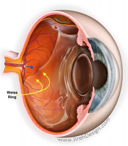
A Weiss ring is the point of attachment between the vitreous and the optic nerve. The optic nerve is round – hence, the Weiss “ring.” Not all patients with a PVD actually have a Weiss ring, but when present is visible upon examination.
A Weiss ring may not necessarily cause cause floaters, but they can. I have had a few patients who complain of a floating ring in their vision.
When I see a Weiss ring in a patient, it is an obvious sign of a posterior vitreous detachment (PVD).
Best Ophthalmic Illustration
This is one of the best illustrations of a PVD, Weiss ring and retinal tear. This illustration was created by Mark Erikson. He is the best ophthalmic illustrator.
The Weiss ring can be seen just in front of the optic nerve where the vitreous was previously attached. The retinal tear almost always, always is anterior to the equator of the eye. In other words, the retinal tear is always in the front half of the retina.
Why? Because anterior vitreous is more adherent to the retina compared to the posterior vitreous.
Most retinal tears are “horseshoe” shaped and have a specific orientation – they are always oriented in the same way due to the adherence of the anterior retina.
If a retinal tear occurs during surgery – tears are usually found in the anterior retina.
PVD and Vitrectomy
Patients with a PVD allow me to most efficiently remove the vitreous. As the vitreous is no longer adherent to the posterior retina, it is much easier to remove free-floating vitreous.
Vitrectomy is much easier to perform with a PVD and done much more efficiently. In patients without a PVD, I prefer inducing a PVD to allow me to remove more of the vitreous…especially vitreous close to the surface of the retina.