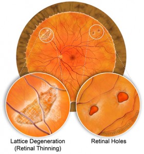
 Lattice degeneration is a common and normal finding in the retina. While it may increase your chance of developing a retinal detachment, it is not a contraindication to vitrectomy (FOV).
Retinal tears and holes associated with lattice degeneration may lead to retinal detachment, but prophylactic laser treatment can prevent or decrease the chances of retinal detachment.
Natural Thinning of the Retina
Lattice degeneration is often described as a natural thinning of the retina and affects about 5-10% of the normal population. It is noted more often in patients with moderate to high myopia.
Atrophic retinal holes are commonly associated with lattice degeneration. These atrophic holes are very similar to taking a pencil erasure and rubbing hard enough until you get a hole in the paper.
Retinal tears may occur with lattice degeneration, too. Both retinal tears and retinal holes may lead to a retinal detachment.
Retinal Detachment
The concern about lattice degeneration is the association with retinal detachments. Either the holes which sometimes are found with lattice degeneration or the tears which occur adjacent to the areas of lattice can cause a rhegmatogenous retinal detachment.
While it is true that patients with lattice degeneration may have an increased risk of retinal detachment, having a vitrectomy performed does NOT increase the risk of retinal tears or retinal detachment anymore than a person without lattice degeneration.
Retinal Tears and Retinal Holes
Retinal tears can happen at anytime. Retinal tears, without a retinal detachment, can usually be treated with laser to prevent the chance of retinal detachment.
Tears and holes are usually treated with laser, or sometimes, cryotherapy (freezing). The only difference is that laser is easier to perform, but not every hole or tear can be treated with laser depending upon location.
Believe it or not, certain tears/holes are difficult to see. For tears or holes which are difficult to see, cryotherapy is an excellent alternative.
Cryotherapy tends to cause more discomfort and swelling.
Retinal Tears During Vitrectomy
It is possible that a retinal tear can be found at the time of vitrectomy (FOV). It may also be possible a retinal tear is created during surgery.
Regardless, both tears and holes found at the time of surgery can be safely treated with laser or cryotherapy.
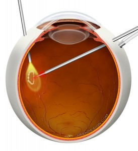
 In the image, a retinal tear is discovered and then treated with a laser probe. In fact, it is standard practice to look for holes and tears at the end of the case. If a tear or hole is found, it most easily treated at this time.
What Does this Mean?
Lattice degeneration is a normal finding of the retina. Patients with pre-existing retinal tears, retinal holes or lattice degeneration may safely undergo FOV, or vitrectomy.